
![]()
|
|
|
Where is your gene expressed?
With Rapid-Scan you'll know in just three hours!
(a complete list of panels and catalog numbers are at the end of this page)
Overview
Rapid-Scan Gene Expression Panels is a system for quickly determining in which tissues a gene is expressed. Rapid-Scan is a PCR-based approach for assessing gene expression that does not require the use of hybridization probes. Each Rapid-Scan panel contains first-stand cDNA from 24 tissues arrayed in 4-log dilution enabling amplification and visualization within the linear range of PCR. Resulting PCR products are then separated in an agarose gel. This makes determination of relative level of gene expression easy. Most commercially-available Northern blots restrict you to analysis of 6 to 8 tissues; Rapid-Scan permits you to survey relative level of gene expression among 24-tissues. Each Rapid-Scan product contains 2 identical 24-tissue panels for analysis of up to 2 separate genes.
Test Results
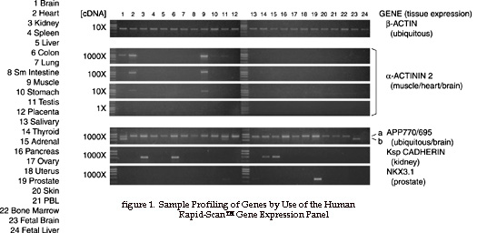
ß-Actin (control) - To facilitate comparison of transcript accumulation in different tissues, the first-strand cDNAs have been normalized using ß-actin as an internal control. Amplification of ß-actin cDNA (using primers provided with the Rapid-Scan panels) shows equivalent distribution in all 24 human tissues (Fig. 1).
a-Actinin - The Rapid-Scan panel was used to determine the relative accumulation of the muscle-specific a-actinin 2 transcript (Fig 1). As expected, a-actinin 2 mRNA was detected predominantly in muscle and heart (muscle; lanes 9 and 2). There was less than a 10-fold difference in mRNA accumulation between the two tissues: at 10X concentration, cDNA levels in muscle were about 2.5-fold higher than that in heart. The a-actinin 2 transcript was not detected in any other tissues other than brain (lane 1), where the a-actinin 2 protein has been shown to anchor the N-methyl-D-asparate (NMDA) receptor on neurons. From a comparison of PCR signals at different cDNA concentrations, it was observed that the a-actinin 2 mRNA level in brain was one-hundredth of that found in muscle.
APP695/APP770 (alternative splicing) - The Rapid-Scan panel may also be used to quantify relative levels of alternatively-spliced transcripts in various tissues or differentiation states. Using a single primer pair, we have compared the expression of two classes of transcripts derived from the amyloid precursor protein (APP) gene (Fig. 1). The APP695 transcript differs from the APP770 transcript by a 225-bp (75 amino acid) deletion, due to alternate RNA splicing. While APP770 was detected in all adult tissues, it was not present in the two fetal tissues (lanes 23 and 24). In contrast, APP695 was detected in fetal tissue but showed greatest accumulation in brain (lane 1). However, trace amounts were found in testis (lane 11) and in other tissues.
AdvantagesRAPID
POWERFUL
EASY
ECONOMICAL
|
|
|
Rapid-Scan Panel Preparation and Quality Control We assembled the Rapid-Scan panels by selecting either 24 frequently-studied human tissues or 24 major mouse tissues and developmental stages. To avoid detection of individual differences in gene expression, we have, whenever possible, pooled tissues from multiple individuals. For the human panel, tissues were from individuals of different ethnicity. For the mouse panel, adult tissues were from outbred Swiss Webster mice, and breast tissues from outbred CD1 mice. Total RNA was isolated and subjected to oligo(dT) selection. The recovered poly(A+) RNA was then examined by Northern blot hybridization, using a ß-actin cDNA probe as control, to confirm RNA integrity. The Poly(A+) RNA was then used to synthesize first-strand cDNA, using oligo(dT) primers and MMLV reverse transcriptase. Individual cDNA pools were confirmed to be free of genomic DNA contamination and to contain complete reverse transcripts of selected rare and long mRNAs, such as those for the transferrin receptor (5 kb) and the ataxia telangiectasia gene (9.4 kb). The first-strand cDNAs from each tissue were then subjected to normalization, such that they all contain an equivalent concentration of ß-actin reverse transcripts. Each cDNA was diluted in water to a series of four concentrations (labeled 1000X, 100X, 10X and 1X), with the lowest concentration (1X) being approximately 1 pg. The diluted cDNAs were subsequently arrayed onto a 96-well PCR plate in the order indicated in Fig. 1. |
|
Rapid-Scan™ Gene Expression Panel |
Catalog Number |
|
Human Rapid-Scan™ Panel (2 Complete Panels*) |
HSCA-101 |
|
Mouse Rapid-Scan™ Panel (2 Complete Panels*) |
MSCB-101 |
|
Drosophila Rapid-Scan™ Panel (2 Complete Panels**) |
DSCC-101 |
|
Human Brain Rapid-Scan™ Panel (2 Complete Panels***) |
BSCD-101 |
*Human and Mouse Rapid-Scan contains 2 identical 24-tissue panels
for analysis of up to 2 separate genes.
**Drosophila Rapid-Scan contains 2 identical 12-discrete
stages/regions of drosophila for analysis of up to 2 separate
genes.
Drosophila Panel is comprised of: Embryo (0-4 hr.), Embryo (4-8 hr.),
Embryo (8-12 hr.), Embryo (12-24 hr.), 1st instar, 2nd instar, 3rd
instar, Pupae, Male head, Female head, Male body, Female body
***Human Brain Panel is comprised of: Frontal lobe, Temporal lobe,
Cerebellum, Hippocampus, Substantia Nigra, Caudate Nucleus, Amygdala,
Thalamus, Hypothalamus, Pons, Medulla, and Spinal cord
![]() Human
Rapid-Scan™ Gene Expression Panel Product Manual in Adobe
PDF format.
Human
Rapid-Scan™ Gene Expression Panel Product Manual in Adobe
PDF format.


